Pathology
Tissue removed at surgery is always sent for histopathology (the examination of tissue under the microscope) examination. In addition very small samples of tissue can be obtained by needle or core biopsy. The microscopic features are assessed by the pathologist and used to determine whether the diagnosis is melanoma. In addition, the pathologist assesses numerous attributes of the melanoma to help determine the stage of the tumour.
The diagnosis of melanoma by a histopathologist can at times be very difficult. Sometimes, specialist histopathologists have different opinions as to whether a given lesion is a benign mole or a malignant melanoma. Second opinions and reviews to confirm the diagnosis is melanoma and also to determine the stage of the tumour are common and can be requested through the Melanoma Unit for a small fee.
There are many features that need to be assessed by the pathologist.
The most important of these include:
- Breslow thickness
- Clark level
- Presence or absence of ulceration
- Mitotic rate
- Evidence of regression
- Invasion of blood vessels, lymphatics or nerves
- Immune cell response
- Excision margins

Pathology Analysis.
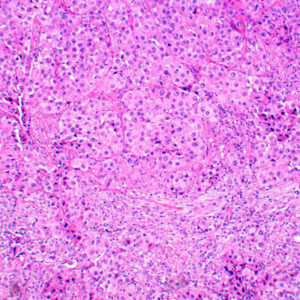
Pathology Slide.