The sentinel lymph node biopsy procedure involves three steps:
- A lymphoscintogram.
- Intraoperative lymphatic mapping with blue dye and a gamma probe.
- Selective biopsy of lymph nodes identified as “sentinel” nodes.
These are explained below.
The sentinel lymph node biopsy procedure involves three steps:
These are explained below.
This is a nuclear medicine scan and is also referred to as a “lymphatic drainage scan” (or a mapping test). This procedure is usually done the day before your operation or the morning before an afternoon operation. You do NOT have to fast (starve) before this test.
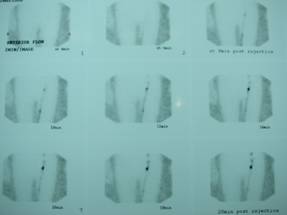
Typical Lymphoscintogram showing a Sentinel Node in the left groin
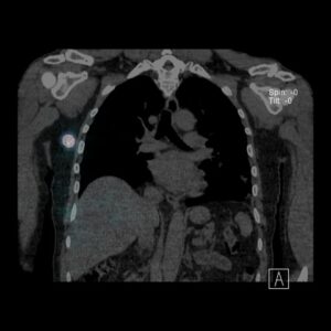
Typical SPECT lymphoscintogram showing a Sentinel Node in the right axilla
The purpose of the test is to accurately identify the location of the lymph nodes that drain the skin around the melanoma. This regional lymph node field might be at risk for containing metastatic disease.
A tiny dose of radioactive tracer is injected into the skin around the site of the primary melanoma. The tracer moves through the skin’s lymphatic channels and special scans are performed to determine the regional lymph node area to which drainage occurs. Scans are done immediately and 2-3 hours later. Although the tracer is radioactive, there is no significant risk to you from its use. Firstly because the dose is so small and secondly because it loses its radioactivity very quickly. The location of lymph nodes identified as “sentinel” nodes – the first nodes on the lymph drainage pathways – will be marked on the skin with indelible ink either in the form of little crosses or tattoos. If crosses have been marked on your skin, please try NOT to wash them off. This test cannot be done as accurately after you have had a wide local excision because the surgery will disrupt the natural lymph drainage pathways from the melanoma site.
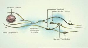
Sentinel node theory
The side effects which may be associated with this scan are slight pain at the injection site during and shortly after the injection – the injections will sting less than the local anaesthetic you had when the melanoma was removed. You may also experience some redness at the injection site for an hour or two afterwards.
This procedure is performed in the operating theatre under a General Anaesthetic. A blue dye called Patent Blue V is injected into the skin around the site of the primary melanoma. The blue dye is rapidly absorbed into the lymphatic channels and moves to the regional lymph nodes. The blue colouring will assist in identifying the sentinel lymph nodes more easily. A Gammaprobe or Geiger counter is also used to identify the “hot” or radioactive sentinel node.
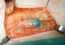
Sentinel node biopsy-blue node
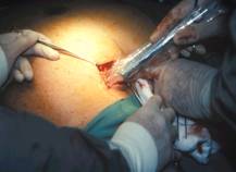
Gammaprobe
This procedure may be accompanied by discolouration of the injected skin and discolouration of the lymphatic channels leaving the injection site. But, this discoloured tissue is normally removed completely as part of the wide local excision procedure. There may be discolouration of the urine lasting no more than 48 hours. There is a possibility of allergic reaction, although this is very rare.
After the intraoperative lymphatic mapping procedure is performed, the sentinel lymph node staging will be done. This consists of removing those lymph nodes which are first in line in the regional lymph node site and therefore the most likely to contain disease if it has spread. The test does not mean the melanoma has spread but simply identifies the at risk nodes.
The sentinel node biopsy is performed in the operating theatre at the same time as the wide local excision (the surgical removal of additional skin and tissue around the site of the primary [original] melanoma). An incision is made in the regional lymph node area(s) identified by the pre-operative lymphoscintigram. Blue and “hot” sentinel node(s) which are identified will be surgically removed and sent to the Pathology Department for examination. If melanoma cells are found to be present in a sentinel lymph node then management has now changed to observation with ultrasound instead of surgery in most cases (depends on size of melanoma deposit). The sentinel node information is essential for accurate staging and potential access to adjuvant treatment drugs (anti-PD1 or BRAF/MEK inhibitors).
The side effects which might accompany a sentinel node biopsy may include the following.